

Eric Betzig, a 2014 recipient of the Nobel Prize in Chemistry and a scientist at Janelia Research Campus, speaks about the latest high-resolution microscopy techniques during a Distinguished Scientist lecture at NCI at Frederick in fall 2015.
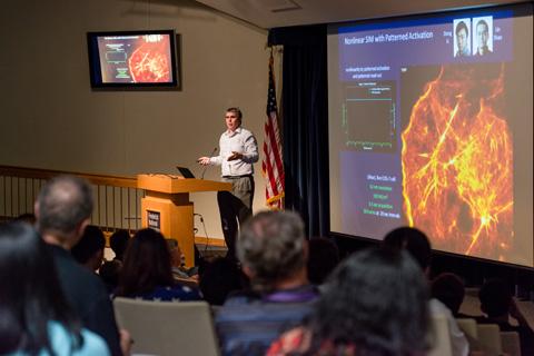
Eric Betzig, Ph.D., a 2014 recipient of the Nobel Prize in Chemistry and a scientist at Janelia Research Campus (JRC), Howard Hughes Medical Institute, in Ashburn, Va., visited NCI at Frederick on Sept. 10 to present a Distinguished Scientist lecture and discuss the latest high-resolution microscopy techniques.
Betzig co-invented photoactivation localization microscopy (PALM) in collaboration with scientists at NIH. PALM achieves 10-fold improvement in spatial resolution of cells, going from the resolution limit of approximately 250 nm in standard optical microscopy down to approximately 20 nm, thus producing a so-called “super-resolution” image. Spatial resolution refers to the clarity of an image or, in other words, the smallest details that can be observed from an image. At 20-nm resolution, researchers can now understand how the cell is constructed at the level of individual proteins. PALM was first published in a landmark Science paper in 2006.
However, Betzig noted that the key limitation of PALM is that it requires several minutes to obtain a super-resolution image, which means the method is too slow to observe the dynamic behavior of sub-cellular organelles and individual proteins in live cells. The speed at which a cellular image can be obtained is referred to as temporal resolution.
To overcome this limitation, Betzig is currently working on microscopy methods that increase spatial resolution while achieving high temporal resolution, in order to capture the rapid molecular movements of organelles and macro-molecules (proteins) in cells that take place at a fraction of a second. The newer microscopy methods are based on structured illumination microscopy (SIM). SIM achieved higher spatial resolution than conventional optical microscopy, but the resolution was not as high as what was achieved using PALM. However, another advantage of SIM is its comparatively lower light dosage, which is well tolerated by live cells. Light dosage is further lowered in some of the newer microscopes by use of a “light-sheet” configuration, where the illumination light is restricted to only the slice of the sample that is being imaged by the camera. This configuration overcomes the problems encountered with older fluorescence microscopy methods, where the entire volume of the sample is continuously exposed to light even though, at any given time, the image is only acquired from a thin slice through the sample.
At the end of his presentation, Betzig noted that the following microscopes in JRC’s Advanced Imaging Center are available to outside users following approval of a research proposal: the lattice light-sheet microscope, the live cell–structured illumination microscope, the widefield multifocal microscope, and the super-resolution 3D iPALM microscope.
Further information about microscopy techniques invented by Betzig can be obtained at the Betzig lab website.
Stephen Lockett, Ph.D., and Jiji Chen, Ph.D., are scientists in the Optical Microscopy and Analysis Laboratory, Leidos Biomedical Research.