

By Nancy Parrish, Staff Writer
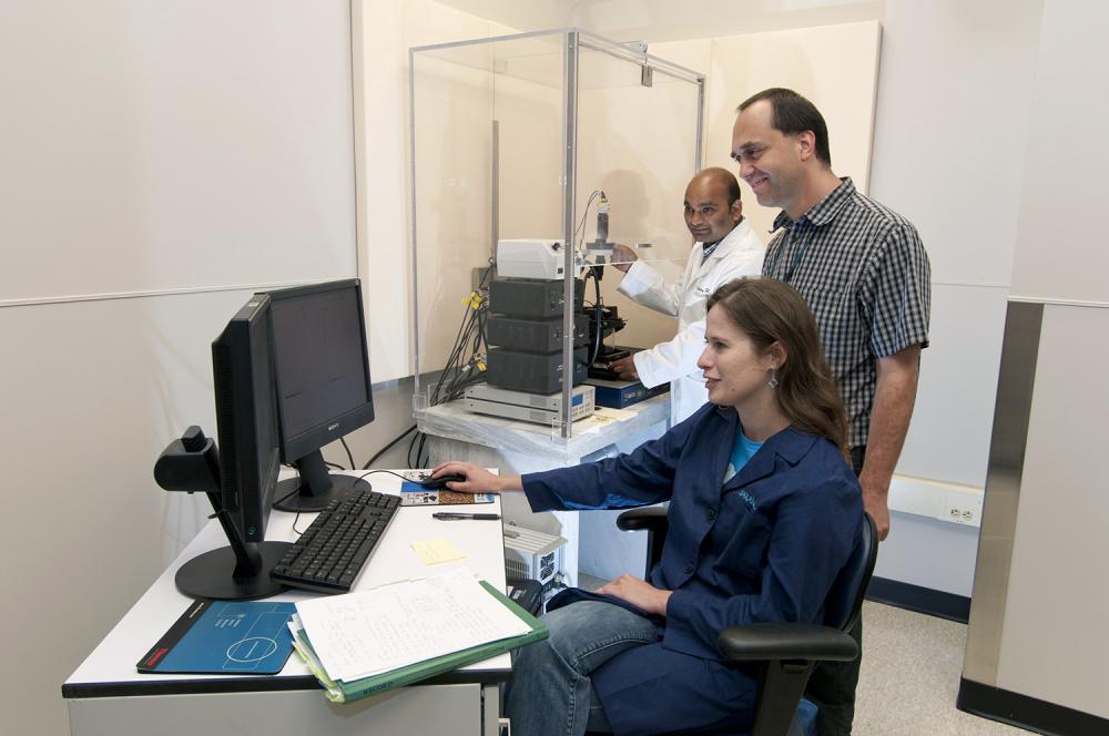
Ulrich Baxa, Ph.D., director of the Electron Microscopy Laboratory (EML), enjoys finally having his staff all in one place.
“Our lab is now all in one location, as compared to our previous situation, with two different locations,” he said. “This will make daily work much easier, in particular for me since I am able to have an office next to the other EML staff.”
A single location is not the only advantage to EML’s relocation to the Advanced Technology Research Facility (ATRF). According to Baxa, the new, larger space offers potential for expansion of staff and technology, which could lead to new partnerships with industry, academia, or other government agencies.
More space has also enabled a better arrangement of the instrumentation. For example, the ultramicrotomes are now located by themselves, in a separate room. These highly sensitive instruments are used almost daily in EML to prepare study samples, some of which may be as thin as 40 nanometers. In the old lab, Baxa said, the ultramicrotomes were in an area where a lot of people walked by, causing vibrations and air movement. These seemingly harmless disturbances can cause slight variations in the section thickness. Having them in their own room reduces the possibility of such interferences and ensures highest quality and consistency of samples, he said.
Baxa’s laboratory works closely with the Nanotechnology Characterization Laboratory (see previous page), which is now across the hall in the ATRF rather than in two other buildings, as it was before the move. Together, these laboratories house powerful microscopes that are used to visualize cellular and subcellular activity, bacteria and viruses, and even a single strand of DNA.
Supporting a Broad Range of Research
Electron microscopy (EM) is frequently used to confirm conclusions derived from other methods, such as a fluorescence (light) microscope, or to make measurements that cannot be done by other, simpler methods (such as, for example, liposome characterization or nanoparticle location in cells), Baxa said.
His laboratory provides significant support to the research of the NCI HIV Drug Resistance Program. “While it is possible to fluorescence-label virus particles in cells, it is not completely straightforward. Electron microscopy of HIV-infected cells, for example, is still the easiest way to study HIV mutations,” Baxa said. Similarly, he noted, “nanoparticles often require EM for characterization and identification in tissue or cell samples.”
EM is also used to check samples for autophagy, the process by which a cell degrades its own components. While autophagy has long been considered a normal cell function, Baxa said, “its importance in cancer?related processes has been recognized recently as it can be a sign of drug toxicity or even a target in developing new cancer treatments.” Autophagy is relatively difficult to study based only on visible light microscopy or fluorescence microscopy, but with EM, it is easily identified in cells, Baxa said.
“About 80 percent of our current work is related to retroviruses, nanoparticles, or autophagy,” Baxa said. The work of his lab has recently supported studies on thyroid regeneration after thyroidectomy and development of novel vaccines against disease, and numerous studies on HIV. “A lot of the projects we support are quite basic research projects that often have broader implications for understanding cancer processes,” he said.
Sources of information in this article: http://www.jic.ac.uk/microscopy/intro_em.html; http://www.nanooze.org/english/articles/article5_powerfulmicroscope.html
The Power of Electron Microscopy
Electron microscopy, known as EM, is critical to biomedical research because it enables researchers to characterize living organisms and analyze cellular behaviors at scales reaching atomic levels. Because it relies on electrons, not light, to produce images, EM is capable of much higher magnifications and generates images in greater detail than those of light-based microscopes.