


Imagine you're building a dresser. You find all the wood and hardware you need and have the instructions, the right tools, and a team of professionals to help. As you and your team put it together, you realize that while the frame is solid, the drawers don't open and close correctly. You might be tempted to feel discouraged by the results.
But a group of scientists from the National Cancer Institute (NCI), Frederick National Laboratory for Cancer Research (FNL), and other collaborating institutions share a different perspective in their recent work published in PNAS. They weren’t trying to assemble furniture but rather generate a new, better mouse model of the most common kidney cancer, clear cell renal cell carcinoma (ccRCC).
After a partially successful attempt, they’re focusing on the lessons learned and how they may be applied to future projects.
The group aimed to develop a model of ccRCC in mice with functional immune systems and the biological conditions that resemble ccRCC in humans, particularly a disabled VHL gene and a resulting accumulation of HIF2 proteins, which can trigger cell overgrowth and tumor formation. Such a model would be useful for testing new ccRCC treatments. While some target the VHL pathway, more effective forms of therapy are needed.
Researchers can use human tumors in specialized mice with suppressed immune systems to test certain drugs, but not immunotherapies, treatments that recruit the immune system to fight cancer. There are no mouse models of ccRCC with intact immune systems in which a combination of treatments including immunotherapies can be tested.
“We need to do better,” said W. Marston Linehan, M.D., chief of the NCI Center for Cancer Research’s Urologic Oncology Branch and a lead author on the study.
So the group used CRISPR/Cas9, a method for gene editing, to prevent specific protein expression for a combination of genes known to be associated with ccRCC, including VHL. Tumor growth in this new mouse model was present after just eight weeks, a much more useful timeframe for drug testing than prior models that hadn’t shown tumor development for over a year and a half. Unfortunately, the tumors didn’t rely on accumulated HIF2 to survive, meaning one of the treatments being tested was ineffective.
It wasn’t what the group aimed for, but they felt they had learned a lot and wanted to share that with the research community.
“We had a story to tell,” said Laura S. Schmidt, Ph.D., principal scientist in the Urologic Oncology Branch, and a first author on the paper, and PNAS agreed.
Relying on Supporting Services
The project was a huge undertaking, with many scientists and complex techniques involved. “This took an enormous effort,” said Linehan. “We’re never going to make progress working by ourselves.”
In addition to the expertise brought together by the collaborating outside institutions, Linehan noted that the team needed to rely on the “invaluable” support services available at FNL.
“It couldn’t have been done anywhere else. It’s painstaking, laborious work that needs really expert people—which we have at the Frederick National Lab,” he said.
One of these experts is Simone Difilippantonio, Ph.D., the director of FNL’s Animal Research Technical Support, which runs over 100 studies per year. Difilippantonio explains that her group handles tasks that range from generating mouse models and performing surgeries and treatments to coordinating each step, ensuring compliance with laws and regulations involving animals, and managing staggered enrollment for various studies.
“If there’s something urgent, we can shift back and forth to accommodate the needs of the study, and everything is very standardized,” she said.
Once the mouse models were established, FNL’s Small Animal Imaging Program (SAIP), under the direction of Joseph Kalen, Ph.D. (recently retired), used magnetic resonance imaging to monitor tumor growth in the mice and their response to the experimental therapies. Another group of experts, FNL’s Molecular Histopathology Laboratory (MHL), helped determine whether the models were similar to human disease via advanced molecular staining and imaging.
MHL fields roughly 3,500 requests per year and is capable of working on about 42 projects at a time, but nevertheless, “quality is the number one priority,” said Baktiar Karim, D.V.M., Ph.D., MHL’s director.
The staff’s experience is the “most important thing” because, under a microscope, “no two cells look the same,” and precision is of utmost importance to each study, Karim said.
For this study, MHL was able to confirm that in many but not all of the mouse models generated, the mouse ccRCC cells had similar form and structure to human ccRCC cells—one of the study’s successes, as it puts the mouse model closer to replicating human cancer.
Though the tumors ultimately were not HIF2 dependent, the team wasn’t deterred. As FNL’s Schmidt put it, “That’s the excitement of the work: there’s always more work to do.”
Turning Failures into Stepping Stones
There’s always more to learn, too. “Cancer is a very complex disease, and we are creating models, but the models are never perfect. We are always learning from these studies; they’re important experiences that lead to the development of the next question,” Difilippantonio said.
To start with, the team presented techniques that other researchers can learn from, including their use of CRISPR/Cas9 gene editing, not often used in mice, to get tumors to grow much faster than previously reported models—useful when planning costly and time-consuming drug studies. And the tumors resembled human tumors, which is important when testing therapeutics that are ultimately aimed toward translation into the cancer clinic, so the team’s process of knocking out several genes can be replicated.
They also learned enough to be able to suggest possible reasons why they weren’t successful in producing HIF2-dependent tumors and to suggest different pathways in which the cancer might function. Researchers could use these findings to generate a successful immunocompetent ccRCC mouse model that could be used for combination drug and immunotherapy testing.
This line of thinking—to apply lessons learned and ask questions that can improve the next study—is what keeps the science moving forward and allows researchers to make progress. Indeed, Schmidt notes that the team’s collaborators at the Dana-Farber Cancer Institute are already working on a slightly modified model based on their findings from this paper—a proverbial new dresser, with new instructions.
“Research is like that—it’s stepping stones. … Sometimes you’re not totally successful at what you tried to do, but you learn things that will help you move forward,” she said.
Image

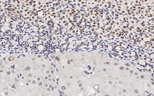
Immunohistochemical staining of tissue sections using a specific antibody to detect Cas9 protein expression. The staining reveals the presence of Cas9 protein in the cancerous cells. Image Credit: Baktiar Karim, Molecular Histopathology Laboratory | Image
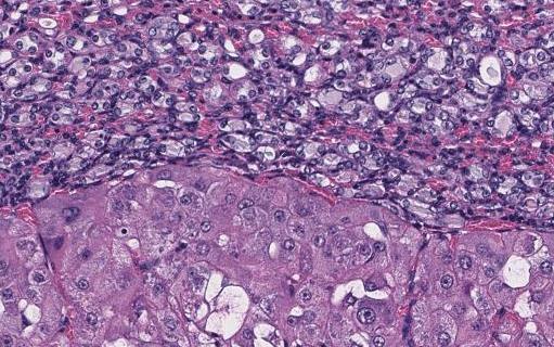
This slide shows tissues treated with the same staining technique as used for image #2, and the results show that the PBRM1 protein is missing from the cancerous cells. Image Credit: Baktiar Karim, Molecular Histopathology Laboratory |
Karolina Wilk is a technical editor in Scientific Publications, Graphics & Media (SPGM), where she writes for NCI Frederick and Frederick National Laboratory’s news outlets and edits scientific manuscripts, corporate documentation, and other writing. SPGM is the facilities’ creative services department and hub for editing, illustration, graphic design, formatting, and multimedia training and support.