

Editor’s note: A version of this article originally appeared on the Frederick National Laboratory website.
Three scientists from the region presented innovative imaging techniques and discussed how they are leveraging them to advance cancer research and ultimately improve human health at the Biotech Connector seminar on Wednesday, May 17, the second in this year’s series sponsored by the Frederick National Laboratory for Cancer Research and the Frederick County Chamber of Commerce.
Optical Microscopy
Anastasiia Aleksandrova, Ph.D., of Leica Microsystems, provided an overview of different contrast methods in optical microscopy that can be used for biomedical imaging, such as cancer-cell imaging.
She focused on fluorescence lifetime and vibrational contrast approaches. Fluorescence techniques, Aleksandrova explained, can image different molecular targets in a specimen, providing precise, but limited information. Fluorescence lifetime techniques provide more functional information about the molecular environment. Vibrational contrast uses the vibrational properties of the molecules’ chemical bonds to visualize them.
“The synergy between all these different types of modalities can provide more information together than each one of them separately,” Aleksandrova said.
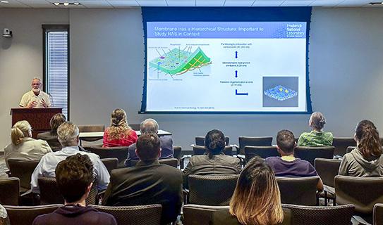
Imaging RAS
Tommy Turbyville, Ph.D., of the RAS Initiative at the Frederick National Laboratory, described the optical microscopy and imaging techniques his team is using to better understand RAS--at a single-molecule level. Mutant RAS is a major driver of cancer; it is found in about 30% of human cancers—making it a critical target for cancer research.
Turbyville described how, using a total internal reflection fluorescence microscope, his team can visualize the RAS proteins in the cell membrane and on synthetic lipid membranes.
“We’ve taken advantage of this [technique] to be able to image RAS at the single-molecule level and then generate maps of how RAS moves laterally in the membrane,” he explained.
With this mapping, researchers can see how RAS behaves in different environments and under different biological conditions and leverage this understanding for small-molecule drug discovery efforts.
Imaging for Cardiovascular Function
To finish the event, Emilia Entcheva, Ph.D., of the Department of Biomedical Engineering at George Washington University, presented about a high-throughput pipeline for manipulating and characterizing human stem-cell-derived cardiomyocytes. Cardiomyocytes are the cells responsible for heart contractions, so this in vitro pipeline can provide important pharmacological information, including cardiotoxicity.
“Any therapeutic that is developed—for cancer, nail fungus, or whatever—is required to undergo cardiotoxicity testing,” Entcheva said. “So, there is a great need to have the right experimental tools to help reduce cost and speed up drug to market.”
Entcheva explained that the pipeline employs both electrical wave (fluorescence) and mechanical wave (dye-free) mapping to detect signals that may indicate cardiotoxicity, and she presented on a proof-of-principle study performed by her laboratory of those techniques.
Future Events
The Biotech Connector is a quarterly series designed to showcase local advances in biomedical research. The next event will be August 31. Join our mailing list to be notified when you can register.
Victoria Brun is a partnership project manager in the Frederick National Laboratory Center for Innovation and Strategic Partnerships, where she provides project management support across the office’s broad portfolio of collaborative projects. Among its functions, CISP establishes the partnerships and collaborations among Frederick National Laboratory scientists and external researchers in government, academia, industry, and the nonprofit research sector.