In 1972, soon after then-President Richard Nixon’s newly established Frederick Cancer Research Center (FCRC) hired its first employees, 24-year-old Kunio Nagashima put on a suit and tie and boarded a Boeing 747 at Tokyo’s Haneda International Airport. An electron microscopist from Kyoto University, Nagashima had a one-way ticket in his hand, bound for the United States and ready to take a new job—sight unseen.
Researchers at the National Cancer Institute (NCI) had embarked upon a massive screening of patient tissue samples in a search for “the human cancer virus,” and they needed scientists trained in electron microscopy to assist with the undertaking. When Nagashima's supervisor in Japan mentioned he had a colleague at NCI who was looking for electron microscopists, "I raised my hand," Nagashima recalled.
After two years at a private contract laboratory then used by NCI, Nagashima came to the FCRC campus as a new Litton Bionetics, Inc., employee and was charged with setting up and running the brand-new Electron Microscopy Laboratory (EML).
A Generous and Willing Collaborator
Over the next 47 years, the FCRC evolved into NCI at Frederick and Frederick National Laboratory, and the EML eventually settled in the Advanced Technology Research Facility in 2010. Despite several laboratory relocations and all the associated upheaval that comes with them—and attending college to earn bachelor’s and master’s degrees while working full-time—Nagashima continued to skillfully assist fellow scientists with research that required transmission electron microscopy (TEM) and scanning electron microscopy (SEM) to capture images of microscopic subjects.
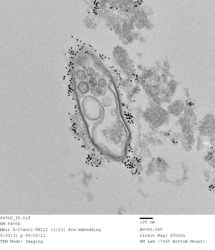
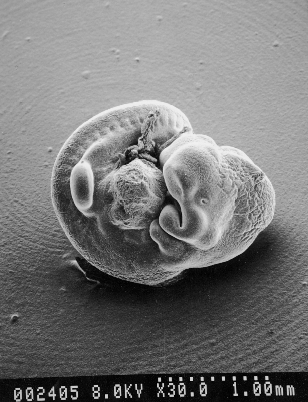
The types of samples brought to the EML for analysis varied greatly: tumor cells, blood cells, viruses, bacteria, patient tissues, animal tissues, insect parts, and more. “We supported over 50 PIs at NCI/NIH and analyzed over 2,000 samples per year,” said Natalia del Val, Ph.D., who directed the EML for several years before leaving the Frederick National Laboratory to become a senior product specialist at Thermo Fisher Scientific.
Nagashima’s meticulous nature and scientific curiosity garnered him a reputation as an insightful collaborator every researcher wanted on their team. “When I joined NCI-Frederick in 1979, Kunio was the resident master of electron microscopy. As my research interests focus on the structure and assembly of virus particles, I quickly came to rely on Kunio for his beautiful work and expert advice,” recalled Alan Rein, Ph.D., senior investigator, HIV Dynamics and Replication Program, Center for Cancer Research (CCR).
Wei-Shau Hu, Ph.D., deputy program director, HIV Dynamics and Replication Program, met Nagashima in the early 2000s, while researching retrovirus assembly. “He was well known as the expert in virus morphology, so I approached him for a collaboration,” said Hu. At the time, Hu was studying a highly charged viral motif, temporarily nicknamed “electric wire,” found in the murine leukemia virus. “Kunio characterized the viral morphology of all the mutant viruses, without which we could not have done the study,” Hu said.
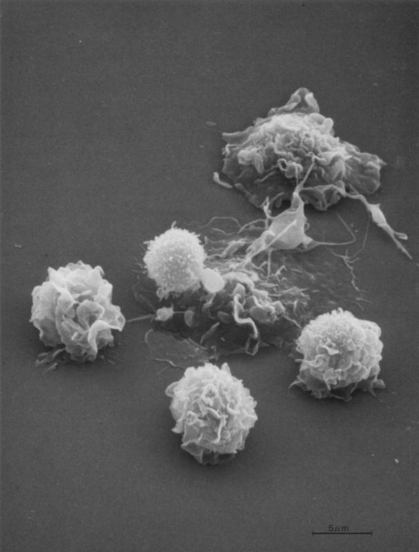
 Nagashima’s colleagues say that no study was too complex or daunting for him. “Whenever I needed to prepare cells for the EM imaging, I always knew whom to ask for help no matter how complicated the conditions were,” said Marina A. Dobrovolskaia, Ph.D., director of Operations and head of the Immunology Section, Nanotechnology Characterization Laboratory. “With Kunio’s help, we learned so many interesting things about nanoparticle interactions with the immune cells.”
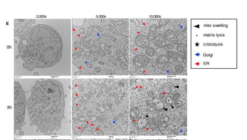
In some cases, Nagashima was more than just an EM guru. His vast experience and knowledge base often led him to have a hand in the design of the experiments themselves. Yoshimi Endo Greer, M.D., Ph.D., associate scientist, Women's Malignancies Branch, CCR, worked with Nagashima to study ONC201, a drug that kills breast cancer cells.
Through his EM analysis, Nagashima found that the drug severely damaged the structure of the mitochondria of the cancer cells. When they collaborated on the design of the next set of experiments, Greer proposed six- and 12-hour timepoints. But Nagashima insisted on adding a three-hour timepoint as well. “It was probably his intuition, but [it] turned out to be such insightful, spot-on advice,” Greer recalled. “With his EM analyses, we confirmed that ONC201 disrupts mitochondria functionally and structurally as early as three hours.”
As a result, they were the first group to report that ONC201 targets mitochondria. Not only did Nagashima’s meticulous work make the discovery possible but “his beautiful EM images were the highlight of the paper,” Greer said.
Mentoring the Next Generation of EM Talent
Nagashima knows the vital role mentors can play in shaping a young person’s career and life path. When Nagashima graduated from high school in Japan, one of his teachers put him in contact with a scientist at Kyoto University who was looking for someone to train in TEM. Nagashima accepted this challenge and found that he had a knack for the work. “Somehow it fit my nature to work on a very small scale,” he said.
Recognizing the value that on-the-job training had on his own career, Nagashima became a dedicated mentor to the next generation of electron microscopists at NCI at Frederick.
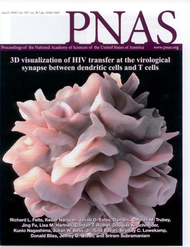
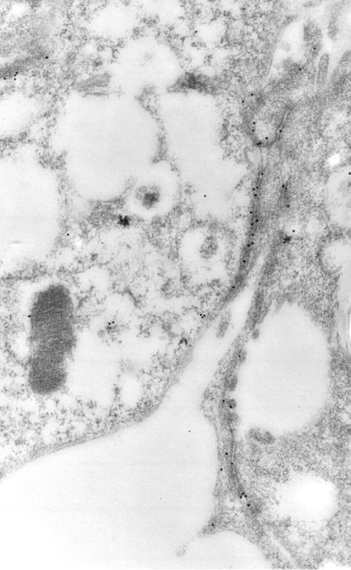
Kedar Narayan, Ph.D., senior scientist and group leader in the Center for Molecular Microscopy (CMM), met Nagashima just after finishing his Ph.D. “I needed to learn the basics of EM sample preparation for my postdoc,” Narayan said. The pair would go on to collaborate on a study of HIV virological synapses. “The end result was two-fold: our paper was accepted as a cover article, and I also realized that my technique would never quite get as good as his!” Narayan added.
Years later, when Narayan was offered the opportunity to lead the CMM, he returned the favor. “I jumped on the opportunity to have Kunio as part of our lab,” he said. At CMM, Narayan said Nagashima became known as the “go-to person for advice, clarifications, and the oft-repeated question ‘Have you seen THIS before?’” Given Nagashima’s unparalleled experience and memory, Narayan said he would usually have an immediate answer.
CMM colleague Adam Harned, electron microscopist and associate scientist, met Nagashima in 1997. “Kunio was my manager for many years when I was a member of the EML, although he acted more like a mentor. He was always more than willing to answer questions no matter how trivial, and he would happily explain how to perform a specific protocol or technique,” said Harned.
Former EML assistant M. Jason de la Cruz is now the manager of the Cryo-EM Facility at Sloan Kettering Institute, Memorial Sloan Kettering Cancer Center in New York, but he fondly recalls his early career as an electron microscopist under Nagashima. “Kunio hired me out of college as an EM technician back in 2003. It was my first real job, and at the time I didn’t think I would stay in the EM field,” de la Cruz said.
But over time and with encouragement from Nagashima, de la Cruz developed an interest in cryogenic electron microscopy (cryo-EM, an extension of traditional transmission electron microscopy), which yields a three-dimensional structure of the viewed object. “I’ve been in the cryo-EM field ever since,” he said.
Passing the Torch
Although his familiar face and congenial manner will no longer be a fixture in the EML, the laboratory that Nagashima so carefully built over nearly five decades will be in good hands upon his retirement. “My crew, they are very skillful people, so I have no concern,” Nagashima said.
One of the “crew” members is Ferri Soheilian, an electron microscopist and associate scientist at the EML who has worked with Nagashima since 2004. “I learned everything to do my job in the field of EM from Kunio. He was, and still is, the master of EM when it comes to technique, methodology, and practices,” Soheilian said.
Christina (Burks) Carpenter, electron microscopist and research associate at the EML, was hired by Nagashima in 2008. “Kunio taught me everything I know about electron microscopy processing and imaging,” she said. “I learned from the best. He is a pleasure to work with, and we shared many laughs over the years.”
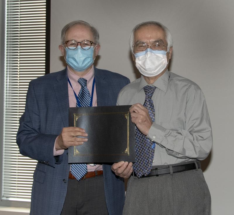
The end of the Nagashima era arrived with the dawn of the new year. Current and former colleagues gathered on January 7 for a COVID-19–safe celebration. Amid the well-wishes and fond memories, Nagashima handed the keys to the EML to Soheilian and Carpenter, who will co-manage the laboratory while a search is held for a new laboratory manager.
“Wish us luck because Kunio truly is irreplaceable!” Carpenter said.
Lisa Simpson is a technical editor in Scientific Publications, Graphics & Media (SPGM), where she edits corporate reports, client projects, and scientific manuscripts, and now writes for the Poster newsletter. SPGM is the creative services department and hub for editing, illustration, graphic design, formatting and multimedia training and support for NCI at Frederick and Frederick National Laboratory.