Ulrich Baxa, Ph.D., and his team are helping to move a microscope the size of a minivan.
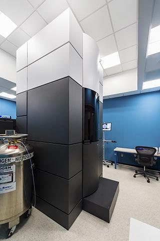
The behemoth, a Titan Krios, is making the 29-mile trek from a Gaithersburg, Maryland, laboratory to its new home at the Frederick National Laboratory for Cancer Research. It’s a highly-sensitive, $7 million device being relocated across a greater metropolitan area with some of the worst traffic in the United States.
“It’s huge amounts of logistics,” said Baxa, the senior microscopist at the National Cryo-Electron Microscopy Facility (NCEF), the group at the national lab that operates the Krios.
It’s nearly impossible to move the Krios in one piece, so the NCEF staff called technicians from Thermo Fisher Scientific, the microscope’s vendor, to disassemble it and package its components in Gaithersburg. A separate team of professional movers transported the parts to Frederick, where the vendor is reassembling it. Baxa and other NCEF staff oversee the project.
The strategy makes transportation easier, but it is not without challenges. Damage to any part of the Krios will delay reassembly, a month-long process that demands perfection. The slightest error could compromise the microscope’s vacuum seal, which is critical for proper function. Even if the assembly seems sound, Baxa and his team won’t know until they test it.
Adding to the challenge, the reassembly is happening in an active scientific facility. Ten feet away, an adjacent room houses a second, functioning Krios that the NCEF uses for high-resolution cryo-electron microscopy (cryo-EM), a method for examining objects and molecules at a near-atomic level.
The relocation is the final step in the NCEF’s effort to consolidate its equipment in one location. The Frederick National Laboratory has been preparing for the move for more than a year. Fortunately, the project hasn’t encountered any major issues so far. The NCEF hopes to perform the first tests on the reassembled Krios in two weeks and have it fully functional before the end of the year.
Then and Now
The NCEF was founded nearly two and a half years ago. Its staff performs cryo-EM for cancer researchers at nonprofit and academic institutions looking to determine molecular structures to aid the drug discovery process.
Collaborators prepare and submit molecular samples, which the NCEF then tests for quality. If the samples pass, they undergo an automated two-day microscopic scan that captures an average of 6,000 images that can be back-projected into 3D structures.
Baxa says the facility has come a long way. The team—which initially comprised him, another microscopist, and a project manager—has doubled in size, adding a third microscopist and two IT specialists. Once the Krios from Gaithersburg is back online, the NCEF will likely hire a fourth microscopist to balance the workload.
The microscopes are another indicator of the facility’s evolution. At its founding, it had only one device, the Gaithersburg Krios. Since then, it added a second Krios and an FEI Glacios transmission electron microscope in Frederick.
Perhaps most importantly, Baxa says the NCEF has dispelled questions, including his own, of whether automated cryo-EM can consistently capture high-resolution molecular structures.
“We knew that it [could] be done, but it hadn’t been done many times before, so it was clear that it was a bit of a challenge,” he said.
Fortunately, the first few projects succeeded—and now, the NCEF has helped more than 100 collaborators. To date, 12 studies using structures obtained at the facility have been published.
Scientific Progress
Baxa takes considerable satisfaction in seeing those studies, even though none lists an NCEF employee as one of the authors. (The facility is credited in the Acknowledgments instead.) They show him that his team is making a difference.
“You read about it, and it’s like, ‘Okay, so what we do has some effect down the line,’” he said.
This validation is especially important because the team is often a few steps removed from its collaborators’ research. Collaborators aren’t required to provide in-depth context for wanting to use the facility’s services, so long as their projects demonstrate a clear connection to cancer or the National Cancer Institute’s mission.
Baxa is particularly proud of a project with scientists at Case Western Reserve University. The NCEF provided several 3D structures of a receptor that stimulates nausea and vomiting in humans, which is helping the Case Western team to develop new drugs that could lessen cancer patients’ nausea during chemotherapy.
Baxa is equally enthusiastic about his team’s overall work and its collaborators’ work. He jokes that he’s becoming something of a technologist because he feels excited when they obtain high-quality data.
“I kind of get proud because we got really good resolution. That tells me our team’s good, our microscope’s good, everything’s good,” he said.
What the Future Holds
The NCEF is expected to continue growing in the coming years, and Baxa hopes to expand its services beyond cryo-EM’s microscopy component. He would like to train collaborators and help them with sample preparation and data analysis, the pre-microscopy and post-microscopy steps in the process.
That growth may come slowly, five or more years in the future. In the meantime, the NCEF will continue to make smaller technical advances and, hopefully, big scientific ones.