

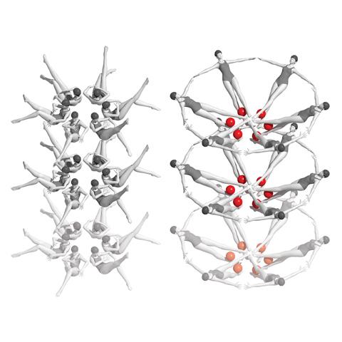
Riboswitches change structure when introduced to biological stimuli, much like synchronized swimmers moving into a new formation.
A recent study at the Department of Energy’s Stanford Linear Accelerator Center National Accelerator Laboratory has literally shed new light on the structural interactions between RNA and another biomolecule.
The researchers, an international team led by the National Cancer Institute at Frederick’s Yun-Xing Wang, Ph.D., senior investigator, Protein-Nucleic Acid Interaction Section, Structural Biophysics Laboratory, Center for Cancer Research, used an X-ray free-electron laser (XFEL) to examine and record how molecules can change the physical structure of RNA.
They aimed the laser at riboswitches, RNA molecules that regulate protein production, on the messenger RNA (mRNA) of Vibrio vulnificus, a multi-drug-resistant bacterium that has a mortality rate of over 50%. The “switch” part occurs when riboswitches change their structure after being introduced to biological stimuli, which in turn alters the mRNA’s protein production functions.
“It has a domino effect on what living things will do, for example, making more proteins or stopping making proteins,” Wang said. Riboswitches may find potential applications in medicine or biotechnology because of their ability to be switched “ON/OFF” to control gene expression.
To study exactly how the change occurs, the researchers encased the Vibrio vulnificus mRNA strands in microcrystals and nanocrystals before introducing them to adenine, a molecule that stimulates the mRNA’s riboswitches and causes a structural change. They then fired a high-speed XFEL, a powerful beam of light that pulses for 5–10 femtoseconds (a femtosecond is one quadrillionth of a second), into the crystals, allowing the team to observe structures and structural changes at different stages of the interaction. The entire interaction process occurs in fractions of a second in solution.
“To get a sense of how long a femtosecond is, let me give you an example: It takes 2.4 seconds for light to travel to the moon and back and 100 femtoseconds to travel through a human hair,” Wang said. “In the femtosecond’s eyes, everything is static! It is basically like someone has a camera with a very, very fast shutter speed that can catch any motion frames.”
Once the crystals are struck by the laser, they diffract the X-rays, which are captured and recorded. The X-rays, in turn, can be used to create high-resolution images of the structures of molecules inside the crystal—snapshots that depict the alterations mere moments apart from each other. The XFEL fires so rapidly that the resulting images can be assembled to form a movie of the process, giving scientists the ability to see the riboswitch changes as they happen in real time.
Previously, most time-resolved studies examined how the molecules reacted when stimulated by light, but, as Wang points out, almost all biomacromolecules interact when trigged by ligands such as adenine instead of light. While technological limitations have hindered the exploration of ligand–molecule interactions in real time, the XFEL has now made it possible to do so.
This study’s success has proven that XFEL technology provides a valuable way to study time-resolved biomolecular interactions. It has also revealed a host of new information about RNA.
Yet the study represents only the preliminary uses of XFELs in scientific research. Because biochemical reactions are often tied to cancer, HIV, and other diseases, researchers using XFELs can study molecular interaction at an atomic level. That opens the door to new ways of understanding those diseases—and hopefully treating them.